Anatomy Of Chest And Ribs / Thorax Chest Anatomy Archives Anatomy Note - Powerful muscles that move the head and arms twelve pairs of ribs extend laterally and anteriorly from the thoracic vertebrae to meet at or near the sternum.
Anatomy Of Chest And Ribs / Thorax Chest Anatomy Archives Anatomy Note - Powerful muscles that move the head and arms twelve pairs of ribs extend laterally and anteriorly from the thoracic vertebrae to meet at or near the sternum.. Chest blunt trauma (cbt) and the resultant rib fractures often lead to thoracic collapse. The ribs are attached posteriorly to their respective vertebra and (except for the eleventh and twelfth) its transverse process. Insert contains images of a typical rib and the first rib. Surface anatomy of anterior chest wall. Anatomy is to physiology as geography is to history:
They also have a role in ventilation; Surface anatomy of anterior chest wall. The ribs are situated one below the other in such a manner that spaces called intercostal spaces are left between them. Paschalides medical publications, 2004, with. The breadth is greater in front than journal of anatomy and physiology.
Thorax Surface Anatomy 4 Edition from doctorlib.info Each rib wraps around the lung and descends approximately 3 to 5 inches. On the standard left lateral chest radiograph, the right ribs are projected behind the left and appear. Posteriorly, the heads of the ribs interdigitate with the vertebrae and are numbered according to the inferior vertebra. How these parts interrelate through joints is described also. The anatomical structure of the 24 ribs in the human body is complex because of the irregular shape and different lengths of each rib. Normal anatomic structures are labeled on posteroanterior (pa) and lateral chest radiographs (figs. The rib cage also anchors the bones of the head, neck, shoulders, and arms to the trunk of the body. Respiratory muscle training online course:
Paschalides medical publications, 2004, with.
Respiratory muscle training online course: Each rib wraps around the lung and descends approximately 3 to 5 inches. ■ identify the basic anatomy seen on a chest radiograph. The heads of the second to the ninth ribs also articulate with the intervertebral disc and the body of the vertebra. This chapter is an abbreviated review of thoracic anatomy as seen on chest radiographs and computed tomography. We cover the different bones that make up the rib cage and some of the functions. Basic rib anatomy consists of a head, neck, tubercle. It discusses the specific anatomy of the ribs and costal cartilages, along with the sternum. The rib cage also anchors the bones of the head, neck, shoulders, and arms to the trunk of the body. To carry out the unique functions performed by. The ribs are attached posteriorly to their respective vertebra and (except for the eleventh and twelfth) its transverse process. We hope you will use this picture in the study and helping chest and abdominal cavities with some organs removed. In some patients an extra joint is seen in the anterior part of the first rib at the point where the bone meets the calcified cartilageneous part (arrow).
Paschalides medical publications, 2004, with. As part of the bony thorax, the ribs protect the internal thoracic organs. It can help you understand our world more detailed and specific. Pathology of the heart, mediastinum, lungs and pleura. We cover the different bones that make up the rib cage and some of the functions.
Chest Wall Anatomy Springerlink from media.springernature.com Continue scrolling to read more below. The breadth is greater in front than journal of anatomy and physiology. Related posts of chest bone anatomy. It can help you understand our world more detailed and specific. It discusses the specific anatomy of the ribs and costal cartilages, along with the sternum. And as you might guess from the word major, it makes up the majority of the chest muscle mass. Posteriorly, the heads of the ribs interdigitate with the vertebrae and are numbered according to the inferior vertebra. Chest blunt trauma (cbt) and the resultant rib fractures often lead to thoracic collapse.
Surface anatomy of anterior chest wall.
The costotransverse ligaments in human: The length of each space corresponds to that of the adjacent ribs and their cartilages; The embryologic and anatomic basis of modern surgery. The ribs are situated one below the other in such a manner that spaces called intercostal spaces are left between them. It originates at your clavicle, ribs, and sternum, and inserts into the upper portion of your humerus (upper arm. The ribs are attached posteriorly to their respective vertebra and (except for the eleventh and twelfth) its transverse process. The purpose of this study was to explore the effect of. Surface anatomy of anterior chest wall. Posteriorly, the heads of the ribs interdigitate with the vertebrae and are numbered according to the inferior vertebra. Learn about chest anatomy with free interactive flashcards. Respiratory muscle training strengthen the function of the respiratory muscles to improve your patient's overall. Spiral ct of thoracic inlet. The bones of the chest and upper back combine to form the strong protective rib cage around the vital thoracic organs such as the heart and.
Chest blunt trauma (cbt) and the resultant rib fractures often lead to thoracic collapse. Each rib wraps around the lung and descends approximately 3 to 5 inches. It discusses the specific anatomy of the ribs and costal cartilages, along with the sternum. Ribs are divided into two basic groups the true ribs consist of 8 ribs, each on the left and right sides of the chest wall. The ribs are situated one below the other in such a manner that spaces called intercostal spaces are left between them.
Thoracic And Abdominal Muscles Lecturio Online Medical Library from d3uigcfkiiww0g.cloudfront.net It originates at your clavicle, ribs, and sternum, and inserts into the upper portion of your humerus (upper arm. We hope you will use this picture in the study and helping chest and abdominal cavities with some organs removed. Moving during chest expansion to enable lung inflation. How these parts interrelate through joints is described also. Related online courses on physioplus. Joints between the ribs and thoracic vertebrae. It discusses the specific anatomy of the ribs and costal cartilages, along with the sternum. The ribs are situated one below the other in such a manner that spaces called intercostal spaces are left between them.
As with all parts of the body, the anatomy and physiology of the chest wall are intimately intertwined.
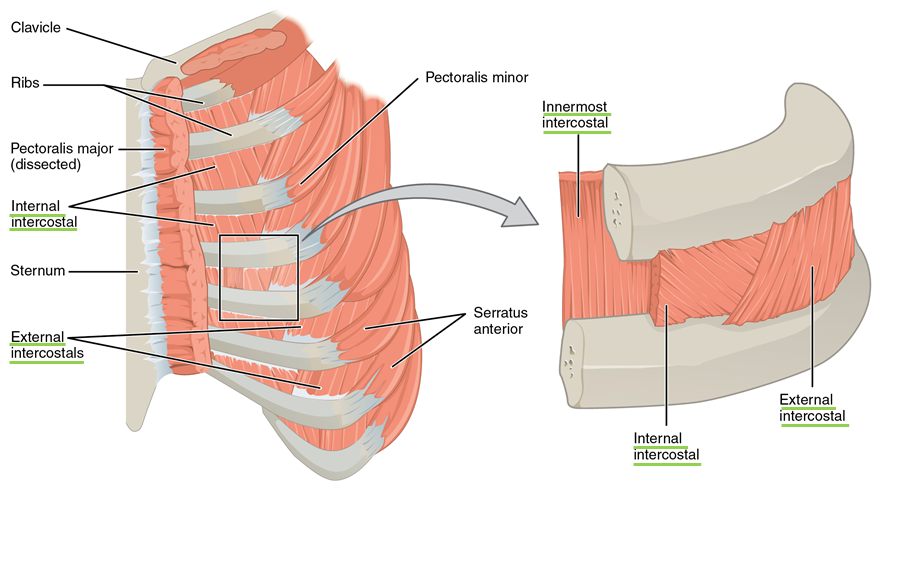
The thoracic rib cage is a diverse structure built for security and support of the underlying organs but is uniquely designed to facilitate respiration. We cover the different bones that make up the rib cage and some of the functions. Respiratory muscle training online course: The chest anatomy includes the pectoralis major, pectoralis minor and the serratus anterior. Surface anatomy of anterior chest wall. Manubrium anteriorly, rib 1 laterally, thoracic vertebrae post… xiphoid process anteriorly, costal cartilages 7 to 10 and rib… The heads of the second to the ninth ribs also articulate with the intervertebral disc and the body of the vertebra. The length of each space corresponds to that of the adjacent ribs and their cartilages; Continue scrolling to read more below. The costotransverse ligaments in human: Bone on hand and foot diagram quiz. Anatomy is to physiology as geography is to history: Normal anatomic structures are labeled on posteroanterior (pa) and lateral chest radiographs (figs.
It describes the theatre of events anatomy of chest. This type of ct scan uses a lower radiation level than a conventional.


0 Komentar